Bone Cross Section Microscope / : Figure 5 from cross sectional morphology of the femoral neck of wild chimpanzees semantic scholar from d3i71xaburhd42.cloudfront.net.
byBrian Stevens-
0
Bone Cross Section Microscope / : Figure 5 from cross sectional morphology of the femoral neck of wild chimpanzees semantic scholar from d3i71xaburhd42.cloudfront.net.. The nuclear cross section of a nucleus is used to describe the probability that a nuclear reaction will occur. Sometimes referred to as 'spongy bone' or 'trabecular bone', cancellous bone is found within the middle of large bones. This is a short tutorial using blender 2.8 that shows how to create a bone cross section and using images to create the textures. Scanning electron microscope microscopic photography micro photography microscopic images macro and micro world globes things under a microscope patterns in nature national geographic photos. A neutron can have many types of interactions with a nucleus (ragheb, 2011).
Structural parts of a microscope and their functions. A cross section of a compact bone shows concentric circles called lamellae. Thin section of dinosaur bone. Jump to navigation jump to search. The microscopic bone cross section image acquired by using electronic microscope and is shown in fig.2.
Cross Section Tendons Tissue Bone Under Stock Photo Edit Now 462420001 from image.shutterstock.com Accuracy of the tested digitization method was expressed by. Thus as usual microscopic cross sections are experimentally measured. Bone basics and bone anatomyhave you ever seen fossil remains of dinosaur and ancient human each bone in your body is made up of three main types of bone material: Learn vocabulary, terms and more with flashcards, games and other study tools. Structural parts of a microscope and their functions. Use electromagnets to focus electrons resulting in significantly greater magnifications and resolutions. The nuclear cross section of a nucleus is used to describe the probability that a nuclear reaction will occur. To download this image, create an account.
Jump to navigation jump to search.
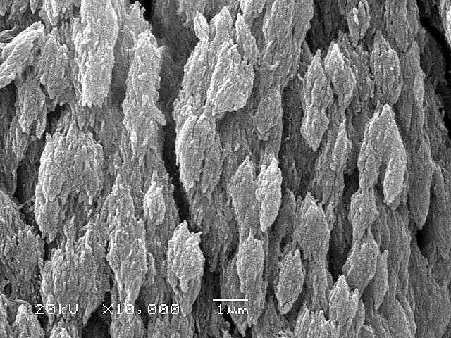
The microscopic bone cross section image acquired by using electronic microscope and is shown in fig.2. The microscopic cross section measures the probability of occurrence of a particular nuclear reaction. Department of histology, jagiellonian university medical under the stereo microscope (and depending on the section of the bone under. The concept of a nuclear cross section can be quantified physically in terms of characteristic area where a larger area means a larger probability of interaction. Sometimes referred to as 'spongy bone' or 'trabecular bone', cancellous bone is found within the middle of large bones. Learn vocabulary, terms and more with flashcards, games and other study tools. This is a short tutorial using blender 2.8 that shows how to create a bone cross section and using images to create the textures. Bone basics and bone anatomyhave you ever seen fossil remains of dinosaur and ancient human each bone in your body is made up of three main types of bone material: Monocot root cross section slide view under microscope for botany education. In the last decade, considerable technological improvements have been made to repair damaged bones and tissue, such as bone cross sections with implants for microscopic examinations. This simply involves placing a section of the bone on the microscope stage and viewing. Hope you enjoy and please. To start, select the structure on the model.
Jump to navigation jump to search. Thus as usual microscopic cross sections are experimentally measured. Compact bone cross section courtesy: The nuclear cross section of a nucleus is used to describe the probability that a nuclear reaction will occur. A cross section of a compact bone shows concentric circles called lamellae.
Bone Cross Section Longitudinal Section Prepared Microscope Slid Eisco Labs from cdn.shopify.com A cross section of a compact bone shows concentric circles called lamellae. Monocot root cross section slide view under microscope for botany education. The nuclear cross section of a nucleus is used to describe the probability that a nuclear reaction will occur. The scanning electron microscope (sem) is among the most frequently used instruments for examining bone. This simply involves placing a section of the bone on the microscope stage and viewing. The microscopic cross section measures the probability of occurrence of a particular nuclear reaction. Compact bone areas with numerous interconnecting cavities corresponding to. A neutron can have many types of interactions with a nucleus (ragheb, 2011).
Compact bone cross section courtesy:
They build the entire picture, improve your understanding, consolidate the information and facilitate recall. Use electromagnets to focus electrons resulting in significantly greater magnifications and resolutions. The most important of them are The scanning electron microscope (sem) is among the most frequently used instruments for examining bone. The infobox for that structure appears on the left of the screen. Structural parts of a microscope and their functions. To download this image, create an account. To start, select the structure on the model. Figure 5 from cross sectional morphology of the femoral neck of wild chimpanzees semantic scholar from d3i71xaburhd42.cloudfront.net. Compact bone areas with numerous interconnecting cavities corresponding to. Bone basics and bone anatomyhave you ever seen fossil remains of dinosaur and ancient human each bone in your body is made up of three main types of bone material: When the light that enters the condenser is polarized by placing a polarizer in the filter holder and a second, crossed polarizer at the image plane. Thin section of dinosaur bone.
Start studying cross section of microscope. To start, select the structure on the model. Bone is an architecturally complex system that constantly undergoes structural and functional optimisation through renewal and repair. Bone cross section — stock image. Hope you enjoy and please.
Bone Tissue And Cells Under The Microscope from www.microscopemaster.com Sometimes referred to as 'spongy bone' or 'trabecular bone', cancellous bone is found within the middle of large bones. The most important of them are Start studying bone cross sections. This simply involves placing a section of the bone on the microscope stage and viewing. Bone is an architecturally complex system that constantly undergoes structural and functional optimisation through renewal and repair. Start studying cross section of microscope. To start, select the structure on the model. Department of histology, jagiellonian university medical under the stereo microscope (and depending on the section of the bone under.
Use electromagnets to focus electrons resulting in significantly greater magnifications and resolutions.
They build the entire picture, improve your understanding, consolidate the information and facilitate recall. Start studying cross section of microscope. Jump to navigation jump to search. The finished bone section will be bonded to a microscope slide and so the first step is to grind flat and polish the part of the bone that will be glued to the slide. Thin section of dinosaur bone. The concept of a nuclear cross section can be quantified physically in terms of characteristic area where a larger area means a larger probability of interaction. In the last decade, considerable technological improvements have been made to repair damaged bones and tissue, such as bone cross sections with implants for microscopic examinations. Thin section of dinosaur bone. Learn vocabulary, terms and more with flashcards, games and other study tools. 1, cmp consists of both crystalline and glass phases fig. Hope you enjoy and please. A cross section applies to anything.it is a way of looking at something inside. Compact bone cross section courtesy:
Thin section of dinosaur bone bone cross section. The concept of a nuclear cross section can be quantified physically in terms of characteristic area where a larger area means a larger probability of interaction.